Overview of Contrast in Fluorescence Microscopy
Pete
Kepf, Certified Vision
Professional- Advanced
Contrast
Machine vision feature extraction for fluorescence
microscopy faces the same challenges as other applications. However,
because of
the low lumen levels involved and the sensitivity required,
signal-to-noise
(S/N) is a chief concern. S/N is affected by the illumination, the
optics and
the dynamic range of the camera.

 We have seen that
illumination consists of emitted light in the form of fluorescence.
Because the
intent is to isolate a specific wavelength to determine the material
properties of a specimen, the less
range of wavelength values seen by the detector, the better the results
will
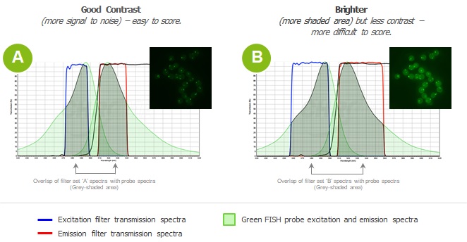
be. Therefore, contrast is more desirable than simply brightness. Figure
2
illustrates two levels of contrast. Both filters pass the desired
wavelength,
but the one on the right also passes much more undesired light. This is
seen as
noise. Better analytical results will be obtained from the left-hand
image due
to higher S/N.
We have seen that
illumination consists of emitted light in the form of fluorescence.
Because the
intent is to isolate a specific wavelength to determine the material
properties of a specimen, the less
range of wavelength values seen by the detector, the better the results
will
be. Therefore, contrast is more desirable than simply brightness. Figure
2
illustrates two levels of contrast. Both filters pass the desired
wavelength,
but the one on the right also passes much more undesired light. This is
seen as
noise. Better analytical results will be obtained from the left-hand
image due
to higher S/N.
Because of their degree of magnification, microscopy
applications have notoriously shallow depths of field. If you took high
school
or college biology, you’ll recall having to frequently adjust the
microscope
lens up and down from the slide to maintain focus. The same laws of
optics
apply to scientific instruments.