 Overview of Focus and Depth of Field in Fluorescence Microscopy
Pete Kepf, Certified Vision Professional- Advanced
Focus and Depth of Field
Focus is defined as that point at which rays of light meet after being refracted or reflected. An object is said to be “in focus” if the rays of light at the detector directly correspond to those at the source. Objects that are not in focus lose definition and noise increases while signal decreases. When this occurs, objects are said to become blurry.
The Depth of Field (DOF) of a lens is its ability to maintain a desired amount of image quality (spatial frequency at a specified contrast), without refocusing, if the object is positioned closer to and farther from best focus (Hollows, 2019). In other words, as an object is positioned nearer or farther from its optimal focus distance, it will begin to blur and contrast will be reduced. This is because the angle between the object and the detector changes with distance and the light rays no longer correspond.
The function of a microscope, of course, is to magnify small images. As a result, fields of view are small as are depts of field. Small changes in position result in large effects from the relocation of the objects viewed. Focusing and refocusing is common on manual microscopes as the user attempts to see “through” the sample or the sample physically moves. We have already seen that reduced contrast results in reduced S/N, so a method that would maintain focus at varying distances would be useful. A common method is termed “Z-stepping” and can be achieved either mechanically or optically.
We are familiar with mechanical Z-stepping from high school biology class. When our amoeba drifted out of focus, we would vary the distance between the lens and the slide to refocus the little critter. The process of moving the lens up or down slightly changed the angle between the lens and the specimen, bringing it back into focus. Optical Z-stepping is similar, except the process is much faster and is performed automatically with a software-adjusted lens.
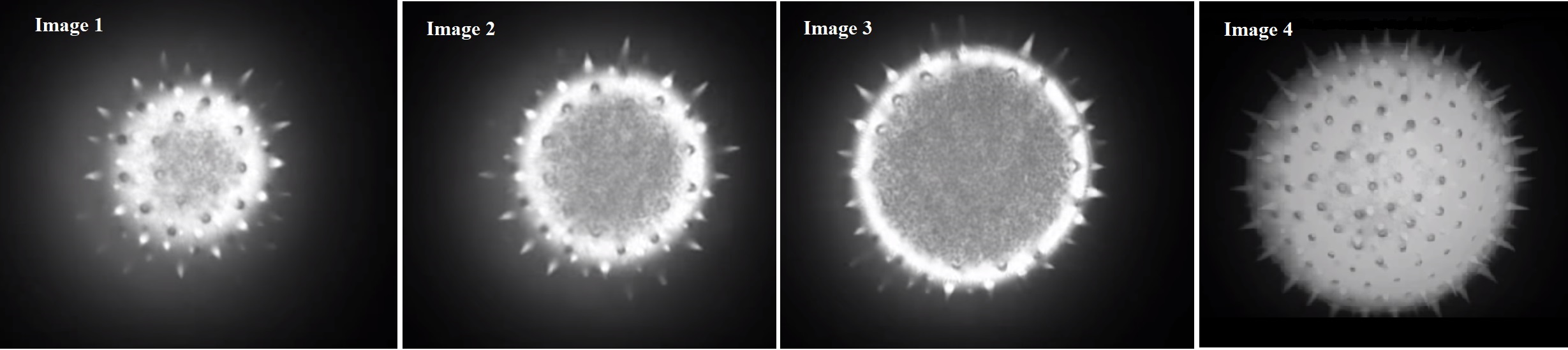
Figures 3-6 illustrate optical Z-stepping. The images in Figures 3, 4 and 5 are acquired at different focal lengths. Although the depth of field is small, a series of images can be acquired very quickly and assembled in software so that the resulting composite image, as seen in Figure 6, shows the complete object in focus.
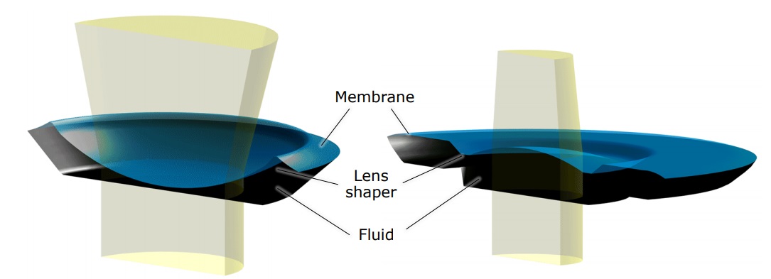
A common method to achieve this is with a liquid lens. A liquid lens consists of a flexible membrane containing a fluid that refracts light just as a glass or plastic lens does. The membrane is expanded and contracted either mechanically or (as in our example) electrically. The result is the ability to vary the focal length of the lens, effectively increasing the depth of field.