Overview of Machine Vision and Fluorescence Microscopy
Pete Kepf, Certified Vision Professional- Advanced
Overview
 Fluorescence microscopy has become an indispensable tool in the biological and material sciences. In biology, it is used to study living cells and tissues, in minerals, it is often used to study coal, and in technology to study ceramics and semiconductors. (Rost, 1999). Fluorescence microscopy is also used in food processing and drug manufacture. Due to its sensitivity, fluorescence microscopy is widely used in the biological sciences. Here we will examine three common machine vision factors and how they are addressed in fluorescence microscopy.
The term “fluorescence” refers to the emission of light at a certain wavelength by an object that is illuminated at a different wavelength. We’re all familiar with a “black light” (ultraviolet light around 400 nanometers [nm]) that causes some materials to emit visible light (green around 540 nm or yellow around 575 nm). Shorter wavelengths have more energy than longer ones. Some of that energy is lost during the fluorescence process, causing the emitted light to have a longer wavelength (different color). Many materials fluoresce when illuminated, or rather “irradiated”. Knowing the fluorescent properties of their molecules has led to an expanding body of fluorescence microscopy applications.
A popular application for fluorescence microscopy is the study of genetic material. On method, termed “FISH” (Fluorescence In Situ Hybridization), uses fluorescent “probes” to optically differentiate different parts of chromosomes according to their emitted wavelength when irradiated. These probes are often derived from fragments of DNA that were isolated, purified, and amplified for use in the Human Genome Project (Wikipedia, 2019), and possess known fluorescence characteristics.
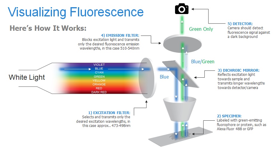
Figure 1 illustrates the fundamentals of fluorescence microscopy as applied to FISH, but actually apply nearly everywhere fluorescence microscopy is applied. White light contains all colors of the rainbow. When white light passes through the excitation filter, only blue is emitted. Blue was previously selected as the wavelength that would cause maximum excitation (fluorescence) of the specimen.
The cyan light strikes a Dichroic (“of two colors”) Mirror. These mirrors are designed to have high reflectivity to one wavelength and high transmissivity to another. They work based on the principle of interference, where one material transmits light (or a wavelength) different from another. We were introduced to interference as children, though we were unaware of it. Remember the multi-rainbow colors on soap bubbles? Soap transmits light differently than air does, resulting in shifting colors as the bubble ingredients, geometry and angles shift. In our FISH example, layers of material that transmit light differently are assembled into a dichroic mirror that transmits green but reflects blue.
The blue light, reflected by the mirror, excites the specimen and it fluoresces green (recall that the emitted wavelength will be longer than that of the initiating light). The green light, and a bit of reflected blue, strike the dichroic mirror. The mirror coating is not perfect, so a bit of blue passes through it also, but is blocked by the green filter so that only the desired wavelength (blue) is seen by the detector.